 Image courtesy of dream designs at FreeDigitalPhotos.net

The purpose of this article is to provide scientific evidence to make the statement that arthritis, spinal discs, joints, and etc may not be causing your pain. I can’t say that it doesn’t increase pain all the time, because there are cases when a disc issue may be promoting inflammation or pressure on a nerve resulting in neurological signs and may need to be surgically mediated.
Please read the Red Flag Section that contains a list of signs and symptoms that may indicate a serious medical condition that requires an appointment with your doctor. There are many cases when surgery may be the only option.
But what if it is not??? That is what this website is about.
There are studies that show people with painful conditions have arthritis and issues with muscles, discs, ligaments, and joints. There are many studies that state this, such as this study published by Cheung and his colleagues here. I can make a long list of articles that state similar comments.
But, what is fascinating is that many studies show that people without any pain also have various issues with muscles, ligaments, joints and discs when they have x-rays, MRIs and CT scans. Many studies have published that there may be minimal cause and effect or relationship of various issues with muscles, bone, joints, discs, and ligaments with pain!
What this means is that we do not know 100% for sure if your pain is related to findings from x-rays, MRIs, and CT scans. So if we assume joint osteoarthritis is causing pain, there is nothing we can do about it. If we assume that there may be other reasons promoting pain, it gives us other options to find what it is. Then there is an opportunity to decrease pain by trying various treatments based on pain science. For example, pain can be related to sensitivity, protective mechanisms, stress, worry, the expectation of pain, changes in the nervous system, the memory of pain, and even changes within the immune system as explained in previous chapters. These components of pain can be treated!
The first time I read about this topic I had a simple question and you may have the same one. Why do people with arthritis tend to have a lot of stiffness and difficulty moving their body?
Arthritic changes may make the body move differently, but it does not mean that it has to be painful. This may be a difficult concept to grasp and I completely understand if it is. It was very difficult for me because at one point, I believed that arthritis always caused pain. What helped me understand this concept was realizing that the belief of something does not make it true.
If we think about the past, most believed that the Earth was flat and if you sailed off to the horizon, you would fall off the edge of the Earth. We now know that this is not true. Many believe that arthritis is causing pain. But there is research evidence indicted that it may not.
Unfortunately, this is not common knowledge and people will not believe it whether it is true for not. So another purpose of this website is to share information that people do not know about, or do not understand in order to help you get out of your pain rut.
Here are some examples of research studies and what they have published:
The Spine:
“The discovery by MRI of bulges or protrusions in people with low back pain may frequently be coincidental.” Published by Jensen MC. 1994
“The relationship between the radiological findings, pain and dysfunction remains unclear.” Published by Ranson CA 2005
“Visually assessed severity of lumbar spinal canal stenosis is paradoxically associated with leg pain and objective walking ability. There is no straightforward association between the stenosis of dural sac and patient symptoms or functional capacity. These findings indicated that dural sac stenosis is not the single key element in the pathophysiology of lumbar spine stenosis” Published by Kuittinen P in 2014
“In typical patients with low back pain (LBP) or radiculopathy, MR imaging does not appear to have measurable value in terms of planning conservative care. Patient knowledge of imaging findings does not alter outcome and is associated with a lesser sense of well-being” Published by Modic MT. 2005
“Lumbar spine radiography in primary care patients with low back pain of at least 6 weeks duration is not associated with improved functioning, severity of pain or overall health status, and is associated with an increase in GP (general practitioner) workload” Published by Kendrick D 2001
“The large proportion of asymptomatic abnormalities (in the lumbar spines of pain free, adolescent, elite tennis players) underlines the poor specificity of these findings, and other sources of pain should be considered.” Published by Ayala F 2006
“Almost half of young Finnish adult aged 21 years had at least one degenerated disc, and a quarter had a bulging disc. Modic changes and disc herniations were, however, relatively rare.” Published by Takatalo J. 2009
Based on a systematic review: “Although there may be an association between degenerative MRI changes and chronic low back pain(CLBP), it is unknown if these estimates accurately represent the association given the quality of included studies, lack of a direct link between degenerative MRI changes and CLBP, and heterogeneity across studies. Thus, a strong recommendation against the routine use of MRI for CLBP evaluation is made. Since there are no data evaluating the efficacy of the surgical treatment of degenerative MRI changes, a strong recommendation is made against the surgical treatment of CLBP based solely upon degenerative MRI changes. Published by Chou D. 2011
“The results of this study suggest that, although some whiplash associated disorders patients are more likely to suffer from long-lasting neck pain, MRI findings cannot explain the symptoms.” Published by Matsumoto M. 2010
“Degenerative changes in the thoracic spine on MRI was observed in approximately half of the asymptomatic subjects” Published by Matsumoto M 2010
“88.5% of subjects in the non-painful group had degenerative changes in cervical discs” Published Okada E. 2011
“Magnetic resonance images of asymptomatic participants revealed abnormalities in 73% of hips, with labral tears being identified in 69% of the joints.” Published by Register B. 2012
“This study documents the high prevalence of anatomical irregularities, including herniation of a disc and deformation of the spinal cord, on the magnetic resonance images of the thoracic spine in asymptomatic individuals” Published by Wood KB 1995
The Upper Extremities:
“Labral tears can occur without symptoms.” Published by Schmitz MR 2012
“The severity of atraumatic rotator cuff tears are not associated with the pain level. Factors associated with pain are comorbidities, lower education level, and race.” Published by Dunn WR 2014
“Structural tendon changes (tendinosis) may be encountered in the symptom free (healthy), normal population” Published by Ustuner E 2013
“Magnetic resonance imaging identified a high prevalence of tears of the rotator cuff in asymptomatic individuals. These tears were increasingly frequent with advancing age and were compatible with normal, painless, functional activity. The results of the present study emphasize the potential hazards of the use of magnetic resonance imaging scans alone as a basis for the determination of operative intervention in the absence of associated clinical findings.” Published by Sher JS 1995
“40% of elite overhead athletes had findings consistent with partial- or full-thickness tears of the rotator cuff. 25% dominant shoulders had magnetic resonance imaging evidence of Bennett’s lesions compared. None of the athletes interviewed 5 years later had any subjective symptoms or had required any evaluation or treatment for shoulder-related problems during the study period.Magnetic resonance imaging alone should not be used as a basis for operative intervention in this patient population.” Published by Connor PM 2003
The Lower Extremities:
“There is evidence of a weak association between joint space width (JSW) of the hip and symptoms, of predictive validity for subsequent joint replacement, and of moderate responsiveness of metric measurement of JSW.” Published by Chu Miow Lin D 2011
“Given the high prevalence of MRI findings in hips of asymptomatic hockey players, it is necessary to cautiously interpret the significance of these findings in association with clinical presentation” Published by Silvis ML 2011
“Knee pain is an imprecise marker of radiographic knee osteoarthritis but this depends on the extent of radiographic views used. Radiographic knee osteoarthritis is likewise an imprecise guide to the likelihood that knee pain or disability will be present. Both associations are affected by the definition of pain used and the nature of the study group. The results of knee x rays should not be used in isolation when assessing individual patients with knee pain.” Published by Bedson J. 2008
“Our results demonstrate that the presence of a plantar calcaneal spur may be an indicator of foot pain independent of plantar fasciitis. Although spurs may not cause foot pain themselves, they may be an indication of other associated conditions.” Published by Moroney PJ. 2014
…
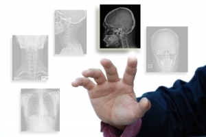
What these research studies have found is that many people without pain have structural issues. So if you have pain and get an x-ray, MRI, or CT scan that shows a structural problem. It does not tell you if the problem found is causing your pain. It may have been there before your injury!
To have even a larger perspective of the relationship of arthritis, discs and degenerative joint disease, the Advanced Physical Therapy Education Institute has created a poster that shares information regarding MRIs, CT scans, and X-rays that can be purchased on their website. Click here to check it out. When the page loads up, you can click on the “larger image” wording under the poster to get an up close look at it. You can purchase one and share with your friends.
This poster makes the point that I am trying to make. Please, please, please give yourself a chance to improve regardless of what a diagnostic test tells you!

To summarize, many research studies have found that diagnostic testing can find issues within your body, but it does not tell us if it is effecting your pain.
If you can remember back 15 years ago when total body scans were popular. It turns out that many research studies have found the lack of benefit of total body scans because of the high rate of false positive findings which led to more unnecessary and invasive tests.
There can also be increased stress and worry that there is a problem leading to increased protective mechanisms and more pain!!
Here are quotes from studies that discusses this exact topic:
“Patient knowledge of imaging findings do not alter outcome and are associated with a lesser sense of well-being.” Published by Ash LM. 2008
“Results suggest that iatrogenic effects (consequence of medical treatment) of early MRI are worse disability and increased medical costs and surgery, unrelated to severity.” Published by Webster BS 2010
“Early MRI without indication has a strong iatrogenic effect (consequence of medical treatment) in acute LBP, regardless of radiculopathy status. Providers and patients should be made aware that when early MRI is not indicated, it provides no benefits, and worse outcomes are likely.” Published by Webster BS 2013
“The American College of Physicians has found strong evidence that routine imaging for low back pain by using radiography or advanced imaging methods is not associated with a clinically meaningful effect on patient outcomes.” Published by Chou R. 2011
“The rate of lumbar spine magnetic resonance imaging in the United States is growing at an alarming rate, despite evidence that it is not accompanied by improved patient outcomes. Over utilization of lumbar imaging in individuals with low back pain correlates with, and likely contributes to, a 2- to 3-fold increase in surgical rates over the last 10 years. Furthermore, a patient’s knowledge of imaging abnormalities can actually decrease self-perception of health and may lead to fear-avoidance and catastrophizing behaviors that may predispose people to chronicity.” Published by Flynn TW. 2011
Here is a blog at this site http://www.bodyinmind.org/spinal-mri-and-back-pain/ that discussed this topic as well. There is a lot of feed back from various health care providers in this blog post.
Here is the kicker. Researchers conducted a study on Aboriginal Australians and explained MRI findings in one group and not in the other. Here is there conclusion:
“Findings are consistent with research in other populations and support that disabling chronic low back pain may be at least partly iatrogenic. This raises concerns for all populations exposed to Western biomedical approaches to examination and management of low back pain. The challenge for healthcare practitioners dealing with people with low back pain from any culture is to communicate in a way that builds positive beliefs about low back pain and its future consequences, enhancing resilience to disability.” (Published by Lin IB in 2013)
The most important point here is to understand that “problems” found in a diagnostic test doesn’t mean that you are doomed and that you will not improve!!! You still have the capability to recover from your pain!
Surgeries are discussed in the next section. So if you have had an unsuccessful surgery, you can still improve!
References:
Photo: http://www.freedigitalphotos.net/images/agree-terms.php?id=10051078
Alyas F, Turner M, Connell D. MRI findings in the lumbar spines of asymptomatic, adolescent, elite tennis players. Br J Sports Med. 2007 Nov;41(11):836-41; discussion 841. Epub 2007 Jul 19. PubMed PMID: 17640926; PubMed Central PMCID: PMC2465278. Free full text
Ash LM, Modic MT, Obuchowski NA, Ross JS, Brant-Zawadzki MN, Grooff PN. Effects of diagnostic information, per se, on patient outcomes in acute radiculopathy and low back pain. AJNR Am J Neuroradiol. 2008 Jun;29(6):1098-103. doi: 10.3174/ajnr.A0999. Epub 2008 May 8. PubMed PMID: 18467522.
Bedson J, Croft PR. The discordance between clinical and radiographic knee osteoarthritis: a systematic search and summary of the literature. BMC Musculoskelet Disord. 2008 Sep 2;9:116. doi: 10.1186/1471-2474-9-116. Review. PubMed PMID: 18764949; PubMed Central PMCID: PMC2542996. Free full text
Canadian Health Services Research Foundation. Myth: Whole body screening is an effective way to detect hidden cancers. Myth Busters. 2009. Available at http://www.cfhi-fcass.ca/Migrated/PDF/11491_newsletter_en.pdf. Accessed July 1, 2014.
Cheung KM, Karppinen J, Chan D, Ho DW, Song YQ, Sham P, Cheah KS, Leong JC, Luk KD. Prevalence and pattern of lumbar magnetic resonance imaging changes in a population study of one thousand forty-three individuals. Spine (Phila Pa 1976). 2009 Apr 20;34(9):934-40. doi: 10.1097/BRS.0b013e3181a01b3f. PubMed PMID: 19532001.
Chou D, Samartzis D, Bellabarba C, Patel A, Luk KD, Kisser JM, Skelly AC. Degenerative magnetic resonance imaging changes in patients with chronic low back pain: a systematic review. Spine (Phila Pa 1976). 2011 Oct 1;36(21 Suppl):S43-53. doi: 10.1097/BRS.0b013e31822ef700. Review. PubMed PMID: 21952189.
Chou R, Qaseem A, Owens DK, Shekelle P; Clinical Guidelines Committee of the American College of Physicians. Diagnostic imaging for low back pain: advice for high-value health care from the American College of Physicians. Ann Intern Med. 2011 Feb 1;154(3):181-9. doi: 10.7326/0003-4819-154-3-201102010-00008. Erratum in: Ann Intern Med. 2012 Jan 3;156(1 Pt 1):71. PubMed PMID: 21282698.
Chu Miow Lin D, Reichmann WM, Gossec L, Losina E, Conaghan PG, Maillefert JF. Validity and responsiveness of radiographic joint space width metric measurement in hip osteoarthritis: a systematic review. Osteoarthritis Cartilage. 2011 May;19(5):543-9. doi: 10.1016/j.joca.2010.12.014. Epub 2011 Mar 23. Review. PubMed PMID: 21396472; PubMed Central PMCID: PMC3268372. Free full text
Connor PM, Banks DM, Tyson AB, Coumas JS, D’Alessandro DF. Magnetic resonance imaging of the asymptomatic shoulder of overhead athletes: a 5-year follow-up study. Am J Sports Med. 2003 Sep-Oct;31(5):724-7. PubMed PMID: 12975193.
Dunn WR, Kuhn JE, Sanders R, An Q, Baumgarten KM, Bishop JY, Brophy RH, Carey JL, Holloway GB, Jones GL, Ma CB, Marx RG, McCarty EC, Poddar SK, Smith MV, Spencer EE, Vidal AF, Wolf BR, Wright RW. Symptoms of pain do not correlate with rotator cuff tear severity: a cross-sectional study of 393 patients with a symptomatic atraumatic full-thickness rotator cuff tear. J Bone Joint Surg Am. 2014 May 21;96(10):793-800. doi: 10.2106/JBJS.L.01304. PubMed PMID: 24875019; PubMed Central PMCID: PMC4018774
Flynn TW, Smith B, Chou R. Appropriate use of diagnostic imaging in low back pain: a reminder that unnecessary imaging may do as much harm as good. J Orthop Sports Phys Ther. 2011 Nov;41(11):838-46. doi: 10.2519/jospt.2011.3618. Epub 2011 Jun 3. Review. PubMed PMID: 21642763.
Jensen MC, Brant-Zawadzki MN, Obuchowski N, Modic MT, Malkasian D, Ross JS. Magnetic resonance imaging of the lumbar spine in people without back pain. N Engl J Med. 1994 Jul 14;331(2):69-73. PubMed PMID: 8208267.
Johal KS, Milner SA. Plantar fasciitis and the calcaneal spur: Fact or fiction? Foot Ankle Surg. 2012 Mar;18(1):39-41. doi: 10.1016/j.fas.2011.03.003. Epub 2011 Apr 13. PubMed PMID: 22326003.
Kendrick D, Fielding K, Bentley E, Miller P, Kerslake R, Pringle M. The role of radiography in primary care patients with low back pain of at least 6 weeks duration: a randomised (unblinded) controlled trial. Health Technol Assess. 2001;5(30):1-69. PubMed PMID: 11701101.
Kuittinen P, Sipola P, Saari T, Aalto TJ, Sinikallio S, Savolainen S, Kröger H, Turunen V, Leinonen V, Airaksinen O. Visually assessed severity of lumbar spinal canal stenosis is paradoxically associated with leg pain and objective walking ability. BMC Musculoskelet Disord. 2014 Oct 16;15:348. doi: 10.1186/1471-2474-15-348. PubMed PMID: 25319184; PubMed Central PMCID: PMC4203914. Free full text
Matsumoto M, Okada E, Ichihara D, Chiba K, Toyama Y, Fujiwara H, Momoshima S, Nishiwaki Y, Hashimoto T, Inoue T, Watanabe M, Takahata T. Prospective ten-year follow-up study comparing patients with whiplash-associated disorders and asymptomatic subjects using magnetic resonance imaging. Spine (Phila Pa 1976). 2010 Aug 15;35(18):1684-90. doi: 10.1097/BRS.0b013e3181c9a8c7. PubMed PMID: 20531071.
Matsumoto M, Okada E, Ichihara D, Watanabe K, Chiba K, Toyama Y, Fujiwara H, Momoshima S, Nishiwaki Y, Hashimoto T, Takahata T. Age-related changes of thoracic and cervical intervertebral discs in asymptomatic subjects. Spine (Phila Pa 1976). 2010 Jun 15;35(14):1359-64. doi: 10.1097/BRS.0b013e3181c17067. PubMed PMID: 20505574.
Modic MT, Obuchowski NA, Ross JS, Brant-Zawadzki MN, Grooff PN, Mazanec DJ, Benzel EC. Acute low back pain and radiculopathy: MR imaging findings and their prognostic role and effect on outcome. Radiology. 2005 Nov;237(2):597-604. PubMed PMID: 16244269.
Moroney PJ, O’Neill BJ, Khan-Bhambro K, O’Flanagan SJ, Keogh P, Kenny PJ. The conundrum of calcaneal spurs: do they matter? Foot Ankle Spec. 2014 Apr;7(2):95-101. doi: 10.1177/1938640013516792. Epub 2013 Dec 30. PubMed PMID: 24379452.
Okada E, Matsumoto M, Fujiwara H, Toyama Y. Disc degeneration of cervical spine on MRI in patients with lumbar disc herniation: comparison study with asymptomatic volunteers. Eur Spine J. 2011 Apr;20(4):585-91. doi: 10.1007/s00586-010-1644-y. Epub 2010 Dec 3. PubMed PMID: 21127918; PubMed Central PMCID: PMC3065617. Free full text
Ranson CA, Kerslake RW, Burnett AF, Batt ME, Abdi S. Magnetic resonance imaging of the lumbar spine in asymptomatic professional fast bowlers in cricket. J Bone Joint Surg Br. 2005 Aug;87(8):1111-6. PubMed PMID: 16049249.
Register B, Pennock AT, Ho CP, Strickland CD, Lawand A, Philippon MJ. Prevalence of abnormal hip findings in asymptomatic participants: a prospective, blinded study. Am J Sports Med. 2012 Dec;40(12):2720-4. doi: 10.1177/0363546512462124. Epub 2012 Oct 25. PubMed PMID: 23104610.
Schmitz MR, Campbell SE, Fajardo RS, Kadrmas WR. Identification of acetabular labral pathological changes in asymptomatic volunteers using optimized, noncontrast 1.5-T magnetic resonance imaging. Am J Sports Med. 2012 Jun;40(6):1337-41. doi: 10.1177/0363546512439991. Epub 2012 Mar 15. PubMed PMID: 22422932. *
Sher JS, Uribe JW, Posada A, Murphy BJ, Zlatkin MB. Abnormal findings on magnetic resonance images of asymptomatic shoulders. J Bone Joint Surg Am. 1995 Jan;77(1):10-5. PubMed PMID: 7822341.
Silvis ML, Mosher TJ, Smetana BS, Chinchilli VM, Flemming DJ, Walker EA, Black KP. High prevalence of pelvic and hip magnetic resonance imaging findings in asymptomatic collegiate and professional hockey players. Am J Sports Med. 2011 Apr;39(4):715-21. doi: 10.1177/0363546510388931. Epub 2011 Jan 13. PubMed PMID: 21233405.
Takatalo J, Karppinen J, Niinimäki J, Taimela S, Näyhä S, Järvelin MR, Kyllönen E, Tervonen O. Prevalence of degenerative imaging findings in lumbar magnetic resonance imaging among young adults. Spine (Phila Pa 1976). 2009 Jul 15;34(16):1716-21. doi: 10.1097/BRS.0b013e3181ac5fec. PubMed PMID: 19770614.
Ustuner E, Toprak U, Baskan B, Oztuna D. Sonographic examination of the common extensor tendon of the forearm at three different locations in the normal asymptomatic population. Surg Radiol Anat. 2013 Sep;35(7):547-52. doi: 10.1007/s00276-. Epub 2013 Feb 17. PubMed PMID: 23417733. *
Webster BS, Cifuentes M. Relationship of early magnetic resonance imaging for work-related acute low back pain with disability and medical utilization outcomes. J Occup Environ Med. 2010 Sep;52(9):900-7. doi: 10.1097/JOM.0b013e3181ef7e53. PubMed PMID: 20798647.
Webster BS, Bauer AZ, Choi Y, Cifuentes M, Pransky GS. Iatrogenic consequences of early magnetic resonance imaging in acute, work-related, disabling low back pain. Spine (Phila Pa 1976). 2013 Oct 15;38(22):1939-46. doi: 10.1097/BRS.0b013e3182a42eb6. PubMed PMID: 23883826.
Wood KB, Garvey TA, Gundry C, Heithoff KB. Magnetic resonance imaging of the thoracic spine. Evaluation of asymptomatic individuals. J Bone Joint Surg Am. 1995 Nov;77(11):1631-8. PubMed PMID: 7593072.

The jobseeker is then expected to upload his resume on the job search engine once
he has selected a job post or opening of a specific company.
Off-Page Optimization: This is a method of optimizing a website where optimization is
done outside the website. If you have a site centered around
dogs then try to get a few inbound links from other dog related sites.
LikeLike
First off I would like to say awesome blog! I had a quick question that I’d like to
ask if you don’t mind. I was interested to find out how you center
yourself and clear your thoughts before writing. I’ve had
a tough time clearing my thoughts in getting
my thoughts out. I do enjoy writing however it just seems like the first 10 to
15 minutes are wasted just trying to figure out how to begin. Any ideas or
tips? Thank you!
LikeLike
These programs are very much helpful for learners all around the world as there are not any restriction based on country or region.
LikeLike
Can A Strong Core Reduce… Even Eliminate Your Joint Pain?
You may have heard that a strong core is important for physical fitness.
Unfortunately, what you’ve heard probably starts and stops with your abs, so you get a “sexy,” ripped” six-pack. That practically guarantees joint pain.
Why?
If your core is weak, your joints compensate by bearing weight that your muscles can’t. Then target one muscle in particular and all the other weak muscles compensate, stressing your joints even more.
Sooner, rather than later, that stress leads to joint pain in a vicious, downward spiral.
What you probably haven’t heard is how a strong core can protect your joints, even end joint pain at the source.
Because your core is a system of 29 pairs of muscles.
But until now, this knowledge has been the exclusive professional secret of the very best sports doctors and physical therapists working for professional sports teams.
Most of us just don’t have access to doctors and physical therapists who know how to teach you to engage, activate and above all, balance your core muscles.
Quickly, without expensive equipment, complicated protocols, or spending several hours a day in physical therapy.
Until now, thanks to former NBA pro and chronic joint pain relief expert Jonathan Bender. Who has a gift for making the complicated. Simple and easy.
If you don’t want to suffer from joint pain any longer… or you want to escape it altogether, click to read “5 Ways Your Core Protects Your Joints.”
It’s already improving my life. I think it can do the same for you.
LikeLike
Great article and very well explained. I really appreciate the insight here in this post and confident it’s going to be helpful to me and many others. Thanks for sharing all the information.
LikeLike